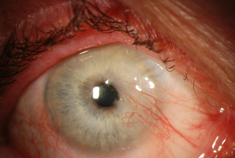
Pterygium Evaluation and Surgery

Pterygium, from the Greek pterygos meaning “wing”, is a common ocular surface lesion originating in the limbal conjunctiva within the palpebral fissure with progressive involvement of the cornea. The lesion occurs more frequently at the nasal limbus than the temporal with a characteristic wing-like appearance.
Etiology
The pathogenesis of pterygia is highly correlated with UV exposure. An increased incidence is noted in latitudes nearer the equator and in individuals with a history of increased UV exposure (outdoor work). Some studies have shown a slightly higher incidence in males than females, which may only reflect a higher rate of UV radiation.
Risk Factors
UV radiation, proximity to the equator, dry climates, outdoor lifestyle.
General Pathology
Histologically, pterygia are an accumulation of degenerated subepithelial tissue which is basophilic with a characteristic slate gray appearance on H&E staining. Vermiform or elastotic degeneration refers to the wavy worm-like appearance of the fibers. Destruction of Bowman layer by fibrovascular ingrowth is typical. The overlying epithelium is usually normal, but may be acanthotic, hyperkeratotic, or even dysplastic and often exhibits areas of goblet cell hyperplasia.
The American Academy of Ophthalmology’s Pathology Atlas contains two virtual microscopy images of tissue samples with Pterygium[2]:
